Zebra medical newsletter June 2026
Dear Zebra Medical community,
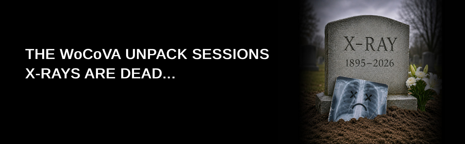
June’s newsletter is another WoCoVA unpack session. And we are focussing on one of the most spoken-about topics at WoCoVA this year: IC-ECG Tip Navigation and the Activated Saline Test (“the Bubble Test”).
European and Australian experts were emphatic that the combination of these two modalities for catheter tip navigation and location represents the new global golden standard. My impression was that the Americans were a tad more non-committal to the X-ray’s demise.
Nevertheless, the most recent literature certainly points towards the adoption of this new standard of care.
The simplicity of both of these tests speaks to the beauty that is our human body:
- The P-wave identification stemming from electrical activity in our hearts.
- The activated saline test being harmless microbubbles that present clearly (albeit quickly) as a “cloud burst” in the right atrium with ultrasound.
- And both tests can be performed quickly, safely and cheaply at a patient’s bedside, in real time, during the procedure.
Imagine. Gone are the days of a static retrospective image after the procedure has been performed. Gone are the days of waiting for the X-ray machine or radiographer. And zero radiation.
Happy reading and always feel free to reach out.
Kind regards,
“… IC-ECG will successfully guide 90-99% of insertions, while the Bubble Test flawlessly covers the remaining minority, meaning patients almost never have to be exposed to the radiation, delays or infection risks associated with X-rays.”
DRS MAURO PITTIRUTI
AND FULVIO PINELLI
IC-ECG Tip Navigation – the New Standard of Care
Chest X-rays have traditionally been considered the gold standard for confirming central venous catheter tip position. Yet as vascular access practice continues to evolve, a growing body of evidence and international expert consensus suggests that routine radiographic confirmation is no longer the safest, most efficient or most cost-effective approach.
Recent WoCoVA 2026 presentations, including experts such as Drs Mauro Pittiruti, Antonio La Greca and Evan Alexandrou, reinforced a clear message: tip location should be confirmed during the procedure, not after it.
Modern technologies such as Intracavitary ECG (IC-ECG) Tip Navigation and the Ultrasound Activated Saline test (“the Bubble Test”) now allow clinicians to accurately identify catheter tip position at the bedside, reducing delays and complications, eliminating unnecessary radiation exposure, and ensuring immediate readiness for therapy.
Today, the GAVeCeLT-SIP Bundle promotes real-time navigation and confirmation, ensuring the catheter reaches the desired location (juncture at the superior vena cava) before the procedure is completed.
How IC-ECG Tip Navigation Works
Intracavitary ECG uses the patient’s cardiac electrical activity to guide catheter placement.
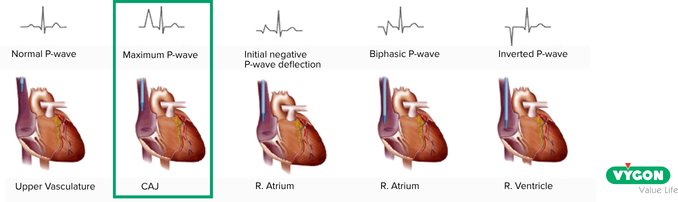
As the catheter advances through the superior vena cava, the P-wave gradually increases in amplitude. The point of maximal P-wave height identifies the cavoatrial junction (CAJ) – the ideal location for most central venous access device tips.
Once the maximal P-wave is identified, the catheter can be secured with confidence, eliminating the need for routine chest X-ray confirmation.
Activated Saline Test (“the Bubble Test”) – the Perfect Complement
For patients where ECG confirmation is not possible – including some patients with atrial fibrillation or altered ECG interpretation – the Activated Saline Test provides an excellent alternative.
How the Activated Saline Test Works
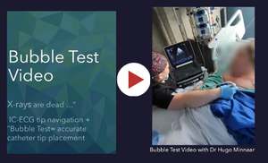
Using transthoracic ultrasound, a rapid saline flush is injected through the catheter while visualising the right atrium. The appearance of microbubbles, seen in 0.5 seconds, confirms the appropriate central tip location.
The technique is fast, radiation-free, and can be performed entirely at the bedside.
WoCoVA Expert Views
Dr Pittiruti described them as a “waste of time, a waste of resources, and potential harm to the patient,” advocating instead for bedside, intra-procedural tip confirmation.
His conclusion was unequivocal: blind insertion followed by post-procedural chest X-ray is no longer acceptable practice.
The future standard of care is real-time tip confirmation using ECG and ultrasound-based technologies, with chest X-rays reserved for only exceptional cases.
Dr La Greca maintained that IC-ECG offers greater accuracy, faster therapy initiation and lower costs than traditional confirmation methods.
His proposed clinical algorithm reserves chest X-rays only for exceptional cases where non-radiologic methods are inconclusive, reinforcing that routine X-ray confirmation is no longer necessary in modern vascular access practice.
Dr Alexandrou reinforced that catheter tip placement reduces complications, thrombosis risk and treatment delays. He highlighted the limitations of traditional confirmation methods, noting that chest X-rays occur after the procedure, while fluoroscopy is costly, resource-intensive, and exposes patients and staff to radiation.
His key message was clear: the value of IC-ECG has been proven. The next step is broader adoption as the standard of care to improve outcomes, increase efficiency and reduce healthcare costs.
The trial Systematic review and meta-analysis on IC ECG in paediatrics and neonates showed savings of AUD $62 per procedure, equating to approximately AUD $100,000 in annual hospital savings. (Zhang et al. 2024)
References:
- Pinelli F, Firenzuoli F, Romagnoli S, Mazzella M, Matarrese D, Bianco E, Gigli V, Villa G. (2026). Intracavitary ECG versus X-ray guidance for central venous access device tip location: A cost-effectiveness analysis. J Vasc Access. 2026 Mar;27(2):519-523. doi: 10.1177/11297298251345989. Epub 2025 Jun 1. PMID: 40452272.
- Brescia, F. et al. (2023). A GAVeCeLT bundle for PICC-port insertion: The SIP-Port protocol. The Journal of Vascular Access.